Category:Virus diagrams
Jump to navigation
Jump to search
Subcategories
This category has the following 6 subcategories, out of 6 total.
A
C
H
I
L
Media in category "Virus diagrams"
The following 125 files are in this category, out of 125 total.
-
 (zh)Virus stucture simple.png 680 × 445; 103 KB
-
 524px-Arenaviridae-esquema.jpg 524 × 599; 66 KB
-
 595768.fig.001.jpg 600 × 557; 164 KB
-
 Adeno-associated viruses-genome.jpg 720 × 540; 12 KB
-
 Adenovirus 3D schematic.png 1,512 × 1,726; 708 KB
-
 Adenovirus NIH.jpg 387 × 469; 23 KB
-
 Adenovirus structure.png 800 × 400; 95 KB
-
 Adenovirus.gif 412 × 493; 20 KB
-
 Adenovirus.jpg 700 × 602; 30 KB
-
 Adenovirus.png 387 × 469; 27 KB
-
 AdenovirusCat.png 387 × 469; 30 KB
-
 Adenoviruses-genome.jpg 2,999 × 2,249; 170 KB
-
 Asfarviridae virion.jpg 189 × 200; 18 KB
-
 CauliflowerMosaicRNA35S.png 857 × 815; 32 KB
-
 Cell with Virus.png 960 × 720; 354 KB
-
 Chronic HBV v2.png 786 × 383; 49 KB
-
 CMVschema-ru.png 721 × 496; 82 KB
-
 CMVschema.ar.jpg 687 × 531; 27 KB
-
 CMVschema.jpg 691 × 463; 104 KB
-
 Conway polyhedron kdktI.png 922 × 914; 78 KB
-
 Coronavirus-HKU1.png 1,868 × 1,498; 3.76 MB
-
 Cowpea chlorotic mottle virus.jpg 320 × 320; 61 KB
-
 Cowpea1.jpg 500 × 440; 110 KB
-
-
 Estructura d'un virus.png 680 × 445; 90 KB
-
 Estructura de un virus.png 1,881 × 1,350; 430 KB
-
 Estructura del VMT.png 374 × 345; 90 KB
-
 Estructura general de un rabdovirus.png 418 × 328; 114 KB
-
 Filo.jpg 500 × 555; 37 KB
-
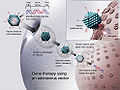 Gene therapy.jpg 495 × 371; 112 KB
-
 HBV genome.png 800 × 791; 179 KB
-
 HCV structure.png 638 × 524; 66 KB
-
 HCV.png 1,200 × 1,200; 1.03 MB
-
 Helical capsid with RNA.png 2,511 × 1,590; 1.54 MB
-
 Helical capsid.jpg 1,802 × 1,352; 104 KB
-
 Hepatitis B virus v2.png 843 × 577; 80 KB
-
 Hepatitis-d-virion-Pathogens-04-00046-g001-1024.png 1,024 × 728; 339 KB
-
 Hexon rus.png 506 × 335; 66 KB
-
 Hexon.ar.jpg 687 × 531; 19 KB
-
 Hexon.png 653 × 496; 60 KB
-
 Hexon1.png 459 × 285; 65 KB
-
 Hexó.png 653 × 496; 65 KB
-
 Hpv16b.gif 1,393 × 1,441; 48 KB
-
 Image-Bacteriophage lambda genome.png 725 × 1,024; 171 KB
-
 Image-vsv.png 701 × 302; 50 KB
-
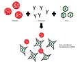 Inhibició de l'hemaglutinació.jpg 960 × 720; 45 KB
-
 Journal.ppat.1007134.g001.tif 3,904 × 2,028; 595 KB
-
 Kapsid-Triangulation-01-fr.jpg 649 × 600; 36 KB
-
 Kapsid-Triangulation-02-fr.jpg 600 × 680; 168 KB
-
 Kapsid-Triangulation-02-fr.png 600 × 680; 415 KB
-
 Kapsid-Triangulation-02.png 1,111 × 1,260; 708 KB
-
 LambdaPhage Genome Linear.jpg 1,214 × 514; 159 KB
-
 Lipu tenpo nanpa jaki - sinpin.svg 326 × 399; 1.09 MB
-
 LMoV-Kapsid.jpg 922 × 2,297; 637 KB
-
 LMoV-Kapsid.png 922 × 2,297; 1.04 MB
-
 Mimivirus 01 FR.jpg 788 × 440; 66 KB
-
 Mimivirus 02 FR.jpg 783 × 595; 91 KB
-
 Mimivirus diagram.gif 169 × 169; 4 KB
-
 Mimivirus.jpg 822 × 624; 96 KB
-
 Mosquito-Carried Diseases (28383412564).jpg 2,503 × 3,716; 4.73 MB
-
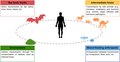
 Mögliche Übertragungswege fledermausspezifischer Pathogene auf den Menschen.tif 3,904 × 2,028; 438 KB
-
 Non enveloped icosahedral virus-es.jpg 404 × 370; 47 KB
-
 Norovirus 2001-2007.png 688 × 408; 20 KB
-
 Nucleocapsids.png 450 × 150; 38 KB
-
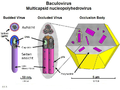 Nucleopolyhedrovirus german.png 800 × 600; 496 KB
-
 Parvo vir.gif 154 × 121; 5 KB
-
 Pathogens-04-00682-g001.png 2,304 × 2,378; 1.25 MB
-
 Peribunyavirus virion structure.gif 1,417 × 780; 337 KB
-
 Phylogeny of Retroviruses.jpg 576 × 400; 38 KB
-
 Polaritaet ambisense nl.png 1,666 × 843; 503 KB
-
 Polaritaet ambisense.png 1,666 × 843; 590 KB
-
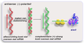
 Polaritaet antisense ar.png 1,666 × 843; 414 KB
-
 Polaritaet antisense nl.png 1,666 × 843; 485 KB
-
 Polaritaet antisense.png 1,666 × 843; 548 KB
-
 Polaritaet sense ar.png 1,666 × 843; 258 KB
-
 Polaritaet sense nl.png 1,666 × 843; 312 KB
-
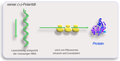 Polaritaet sense.png 1,666 × 843; 355 KB
-
 Poliovirus binding receptor 1DGI.png 800 × 911; 1.02 MB
-
 Prophage-ar.JPG 392 × 425; 29 KB
-
 Prophage.JPG 392 × 425; 28 KB
-
 PSTviroid.png 1,184 × 86; 5 KB
-
 Reassortment HiRes.jpg 2,159 × 3,688; 2.77 MB
-
 Retroviren im Primaten-Genom (german).png 567 × 473; 104 KB
-
 Retrovírus.png 172 × 153; 10 KB
-
 Sarampo2.tif 1,317 × 1,125; 177 KB
-
 Sarampo3.tif 1,524 × 1,295; 185 KB
-
 Serotype.png 800 × 600; 102 KB
-
 Sistèma immunitari - Memòria immunitària de tèrme lòng.png 863 × 605; 72 KB
-
 Structure of a bacteriophage.jpg 1,500 × 1,500; 222 KB
-
 Stup Virus.png 500 × 500; 20 KB
-
 Symmetrieachsen Kapsid.png 1,476 × 591; 181 KB
-
 Tipus Virus.png 460 × 270; 51 KB
-
 TMV structure full.png 2,638 × 1,417; 1.15 MB
-
 TMV structure simple gl.png 800 × 570; 275 KB
-
 TMV structure simple-es.png 640 × 456; 256 KB
-
 TMV structure simple.png 1,474 × 1,050; 177 KB
-
 TMV Structure zh.png 374 × 345; 84 KB
-
 TMV Structure-ar.png 374 × 345; 98 KB
-
 TMV Structure.png 374 × 345; 81 KB
-
 Tobacco mosaic virus structure.png 720 × 360; 86 KB
-
 Tobacco mosaic virus tmv2.png 1,024 × 1,748; 1.28 MB
-
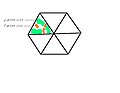
 Triangulationszahl3.png 526 × 239; 30 KB
-
 Viirus.png 156 × 146; 9 KB
-
 Virion.png 600 × 550; 164 KB
-
 Virus capsid T number.tif 1,992 × 1,484; 494 KB
-
 Virus nanometers diameter compared with a cellule.JPG 1,274 × 748; 47 KB
-
 Virus scheme.jpg 1,754 × 3,508; 346 KB
-
 Virus structure simple.png 680 × 445; 82 KB
-
 Virus tegument.JPG 317 × 184; 20 KB
-
 Virus-Budding-Typen.jpg 2,067 × 752; 432 KB
-
 Virus-Budding-Typen.png 2,067 × 752; 531 KB
-
 Virus-Envelope-Dimer.jpg 1,890 × 1,890; 844 KB
-
 Virus-Envelope-Dimer.png 1,890 × 1,890; 1.56 MB
-
 Virus-types-vi.png 460 × 270; 30 KB
-
 Virus-types.png 460 × 270; 32 KB
-
 Virus-types3.png 517 × 304; 125 KB
-
 Viruses-12-00784-g001.png 3,358 × 1,640; 416 KB
-
 Viruses-13-00435-g001-A.png 1,267 × 1,082; 1,007 KB
-
 Viruses-13-00435-g001-v2.png 948 × 645; 471 KB
-
 Viruses-13-00435-g004-A.png 2,926 × 1,647; 694 KB
-
 Viruses-13-00435-g004-B.png 2,926 × 2,050; 1.03 MB
-
 Viruses-Entering-Cell.png 1,000 × 1,000; 248 KB
-
 Vis2.jpg 1,920 × 1,819; 405 KB
-
 Vírusfajták.png 460 × 270; 66 KB
-
 Yutin 2013 bmc virophage polinton transpoviron.png 1,200 × 723; 468 KB
Virus_stucture_simple.png)


.jpg)


.png)


