File:Embryo in flower.png
Jump to navigation
Jump to search

Size of this preview: 598 × 599 pixels. Other resolutions: 240 × 240 pixels | 479 × 480 pixels | 766 × 768 pixels | 1,022 × 1,024 pixels | 2,044 × 2,048 pixels | 3,000 × 3,006 pixels.
Original file (3,000 × 3,006 pixels, file size: 2.97 MB, MIME type: image/png)
Captions
Captions
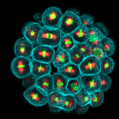
Cell divisions of a sea urchin embryo
Summary
[edit]
| Description |
English: This is a blastula-stage sea urchin embryo. In turquoise we see the membranes of dividing cells, in red the microtubules of the mitotic spindle that act as "ropes" to pull the chromosomes, and in green the DNA in the form of chromosomes. To manage various vital functions, each organ is protected by an epithelium, whose architecture is essential for its barrier function. Cell division and its precise orientation are essential for maintaining the integrity of these tissues. Dysregulation of cell division orientation is linked to the emergence of epithelial cancers. There are many players involved in regulating orientation: microtubules, cell shape and several membrane proteins. But how all these players work together remains a mystery. To study the orientation of division in epithelia, I use an atypical model: the blastula of the sea urchin embryo. This project provides new tools for studying cell division, with numerous implications for the biology of epithelial cancers. The technology employed is confocal microscopy. Model LSM980 in AiryScan mode. x63Oil objective. Image subsequently colored with Fiji.
Français : Ceci est un embryon d'oursin à stade blastula. En turquoise nous voyons les membranes des cellules en division, en rouge les microtubules du fuseau mitotique qui servent de "cordes" pour tirer les chromosomes, et en vert l'ADN sous forme de chromosomes. Pour gérer différentes fonctions vitales, chaque organe est protégé d’un épithélium, dont l’architecture est essentielle pour sa fonction de barrière. La division cellulaire et son orientation précise sont essentielles pour maintenir l’intégrité de ces tissus. Des dérégulations de l’orientation de la division sont liées à l’émergence de cancers épithéliaux. Les acteurs qui régulent l’orientation sont nombreux : les microtubules, la forme des cellules et plusieurs protéines membranaires. Mais comment tous ces acteurs coopèrent ensemble reste un mystère. Pour étudier l’orientation de la division dans les épithéliums ; j’utilise un modèle atypique : la blastula de l’embryon d’oursin. Ce projet apporte de nouveaux outils pour étudier la division cellulaire, avec de nombreuses répercussions sur la biologie des cancers épithéliaux. |
| Date | |
| Source | Own work |
| Author | AudeNommick |
Licensing
[edit]
I, the copyright holder of this work, hereby publish it under the following license:
This file is licensed under the Creative Commons Attribution-Share Alike 4.0 International license.
- You are free:
- to share – to copy, distribute and transmit the work
- to remix – to adapt the work
- Under the following conditions:
- attribution – You must give appropriate credit, provide a link to the license, and indicate if changes were made. You may do so in any reasonable manner, but not in any way that suggests the licensor endorses you or your use.
- share alike – If you remix, transform, or build upon the material, you must distribute your contributions under the same or compatible license as the original.
| This file was uploaded as part of Wiki Science Competition 2023. |
File history
Click on a date/time to view the file as it appeared at that time.
| Date/Time | Thumbnail | Dimensions | User | Comment | |
|---|---|---|---|---|---|
| current | 13:56, 5 December 2023 | | 3,000 × 3,006 (2.97 MB) | AudeNommick (talk | contribs) | Uploaded own work with UploadWizard |
You cannot overwrite this file.
File usage on Commons
The following 7 pages use this file:
- User:FredD/Echinoderm news/2023/12/1–15
- User:FredD/Echinoderm news/2023 December 1-15
- User:Kruusamägi/Teadusfoto2
- Commons:Wiki Science Competition 2023
- Commons:Wiki Science Competition 2023/Images
- Commons:Wiki Science Competition 2023/Microscopy images
- Commons:Wiki Science Competition 2023/Winners/France
File usage on other wikis
The following other wikis use this file:
- Usage on en.wikipedia.org
- Usage on nl.wikipedia.org